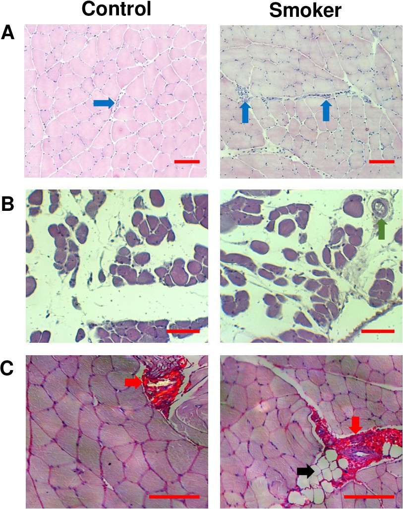
Fig. 1. Representative photomicrographs for the control and smoker groups stained with hematoxylin and eosin (A and B) and picrosirius red (C) showing: (A) inflammatory cells; (B) blood vessels; and (C) collagen and fat deposition. Blue arrows: inflammatory cells. Green arrow: blood vessel. Red arrows: collagen deposit. Black arrow: adipocyte deposition. Scale bars = 50 μm.